Microscopes are standard equipment in limnology, as the species identification of most organisms found in freshwater is not possible without optical aids. Good light microscopes are essential for the identification of larger invertebrates,
but the specialization of the company in the taxonomic study of aquatic dipterans, and in particular the family Chironomidae, required the purchase of high-resolution light microscopes and the employment of different methods for a better visualizing, such as phase contrast and interference contrast.
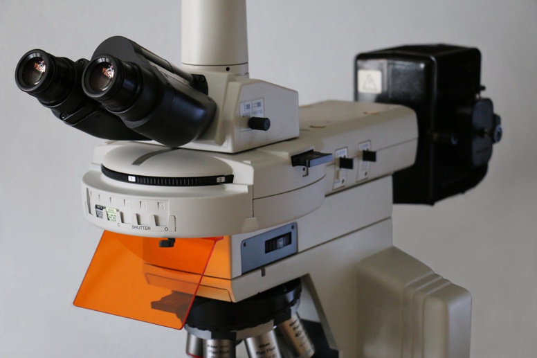
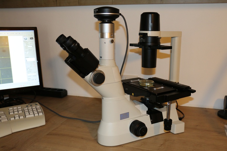
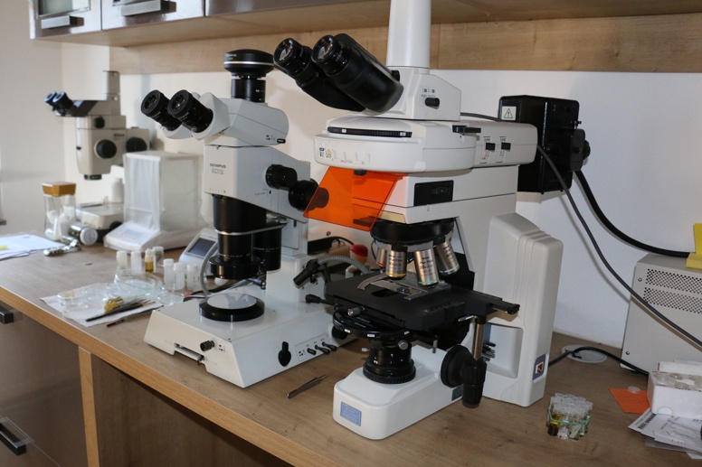
The Eutaxa office owns a range of optical equipment for taxonomic processing, scientific documentation and digital analysis of specimens and samples (measurement, counting of particles, etc.). The epi-fluorescence equipment (top left) and an inverted microscope (top) are among the latest acquisitions.
Light Microscope & Binoculars
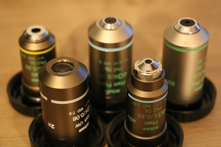
The light microscopes are equipped with plan-fluor and plan-apochromat objectives with magnification factors of 2x to 100x. Various methods are available for the visualization of weakly contrasting structures, such as dark field, phase contrast, polarization, interference contrast and epi-fluorescence.
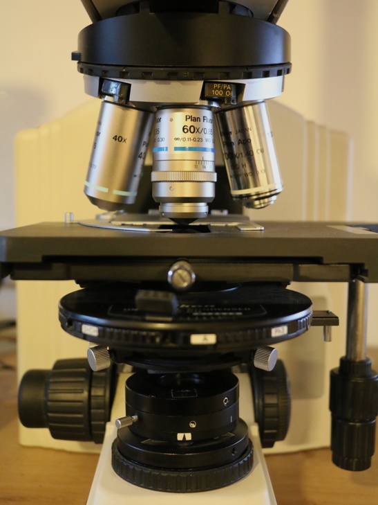
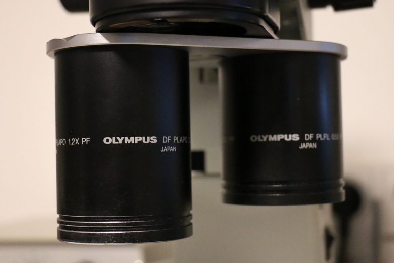
Several binocular devices are available, which are equipped with different types of objective lenses. The range extends from low magnification objectives for the documentation of larger specimens (like juvenile fish or big invertebrates),
to high-magnification, high-resolution interchangeable plan-apo objectives, which enable the imaging of small specimens with transmitted and/or reflected light as well as the taxo-nomic processing of small organisms (e.g. larvae from the dipteran family Chironomidae).
Micro-photography
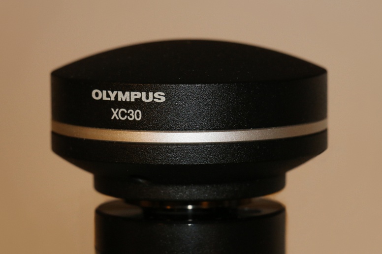
Almost all microscopes are equipped with microcamera adaptions and corresponding facilities for photomicro-graphy. The camera used for imaging is an XC30 digital color camera with a resolution of 2080 x 1544 pixels (top left image). A lower resolution of 1040 x 772 pixels is usually selected as the standard for reproducing images in electronic media (e.g. for the digital identification keys of the Eutaxa series).
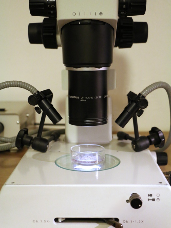
Picture above: Laboratory: Microscope department with two binoculars and a large light microscope.
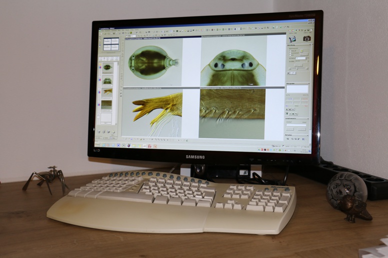
A later version of the Analysis software is used for recording and the calculation of images with an unlimited depth of focus (extended focal imaging) (bottom left image). Due to compatibility problems with newer operating systems, a switch to newer image analysis software is planned.
The Analysis software is not only used for image production, but also for particle counting or for length and area calculations and thus for taxonomic processing of samples.
older devices
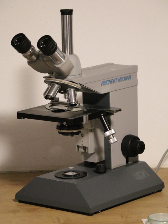
In addition to the more microscopes, the collection also includes some older devices with less high-resolution objectives. These include two microscopes from the Reichert company, a Neovar laboratory microscope (pictured right) from the student days and a Zetopan-Pol for polarization and reflected light microscopy (pictured right, center) acquired from an estate, including extensive accessories. This device is no longer in use, but it is a technical masterpiece and impresses with its fine mechanical workmanship and with numerous details.
outdoor devices
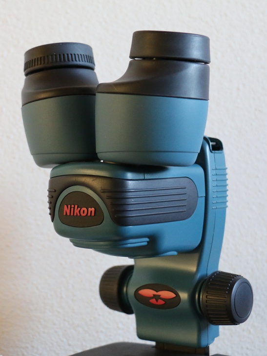
A portable field binocular with battery-powered illumination and plan apochromat objective is used to check invertebrat samples taken in the field. This device was frequently used, especially on excursions abroad in the Mediterranean region (Northern Italy and Peloponnese).